3D-SiSP for 3D multi-spheroid quantitative analysis
Scientific Reports publication on 3D multi-spheroid CSC models and drug response profiling
High-content confocal analysis of tumorigenesis, cancer stem cells, and drug response in 3D cholangiocarcinoma cultures
📖 Brief introduction
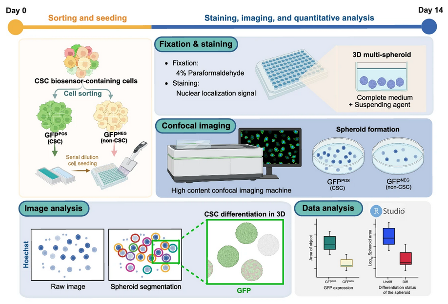
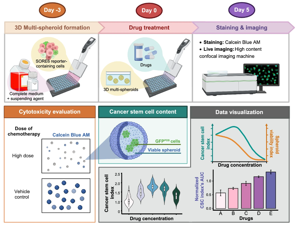
3D Surface Integrative Spheroid Profiling (3D-SiSP) is a high-content confocal imaging–based methodology developed for quantitative analysis of 3D multi-spheroid cultures in cholangiocarcinoma (CCA).
This approach was designed to overcome the limitations of traditional 2D cultures and length-based spheroid measurements by providing a flexible and scalable framework that captures both spheroid morphology and cancer stem cell (CSC) dynamics.
On this page, we provide two key resources linked to our Scientific Reports (2025) publication (Kongtanawanich et al., 2025):
- Wet-lab protocols: step-by-step laboratory methods published on protocols.io, covering spheroid preparation, CSC biosensor use, tumorigenicity assays, and drug-response evaluation.
- R coding tutorials: five analysis modules that guide users through quantitative image analysis, CSC differentiation assessment, and drug-response profiling in 3D cultures.
Together, these resources allow researchers to reproduce the experiments, adapt the pipelines to their own 3D culture systems, and integrate both experimental and computational workflows for CSC research and anti-cancer drug testing.

🧪 Wet-Lab Protocols
The protocols were published in protocol.io in the colletion name 3D-SiSP: High-content confocal analysis of tumorigenesis, cancer stem cells, and drug response in 3D cholangiocarcinoma cultures


🚀 R Coding Tutorial
⚙️ Installation requirements
R (≥ 4.3.3) and RStudio
See R installation guide.
🔬 Analysis Modules
The analysis tutorial is composed of 5 coding modules:
- Evaluating the in vitro tumorigenicity of CSC candidate for the 3D multi-spheroid model by determining spheroid number using 3D-SiSP analytical method.:
-
Evaluating the in vitro tumorigenicity of CSC candidate for the 3D multi-spheroid model by determining object area using 3D-SiSP analytical method.:
- Investigating the relationship between spheroid size and their differentiation status for the 3D multi-spheroid model using 3D-SiSP analytical method.:
- Evaluating CSC content and cytotoxicity under anti-cancer drug treatments for the 3D multi-spheroid model using 3D-SiSP analytical method.:
- Comparing CSC content among cytotoxic drugs across a range of spheroid viability index.:
🔗 Related Projects and Resources
- Cholangiocarcinoma Stem Cell ResearchProject page →
- Computational Biology Portfolio
- Phenomics – Coding × High-Throughput Imaging (2D/3D Models) Project page →
- The 3D multi-spheroid model was central to both (Kongtanawanich et al., 2024), (Kongtanawanich et al., 2025) and (Kongtanawanich et al., 2025), with intensive quantitative analysis showcased in the latter.
- This project also empowers wet-lab data from three models (2D, 3D multi-spheroid, and 3D multicellular spheroid) by integrating dry-lab coding pipelines to generate deeper insights into cancer biology.
- Phenomics – Coding × High-Throughput Imaging (2D/3D Models) Project page →
🧮 Citation
If you use 3D-SiSP in your research, please cite:
Kongtanawanich, K., Jamnongsong, S., Hokland, M. et al. High-content confocal analysis of tumorigenesis, cancer stem cells, and drug response in 3D cholangiocarcinoma cultures. Sci Rep 15, 31387 (2025). DOI: 10.1038/s41598-025-16144-9
👩🔬 Contributors
This project is developed and maintained by
Namthip Krittiyabhorn Kongtanawanich (@KuchikiNamthip) at the Siriraj Center of Research Excellence for Precision Medicine and Systems Pharmacology, Faculty of Medicine Siriraj Hospital, Mahidol University, Bangkok, Thailand:contentReference.


